Blood oxygen saturation (SpO2), as the name suggests, refers to the oxygen concentration in the blood in the peripheral arteries. It is another key physiological parameter reflecting the respiratory and circulatory functions of the human body, following heart rate, blood pressure, respiratory rate and body temperature.
The finger clip type oximeter commonly used now uses optical principles to detect blood oxygen saturation. The main principle is to use the spectrum of hemoglobin to obtain approximate blood oxygen data. More detailed data requires further experimental testing.
In fact, there is very little O2 that exists directly in the blood in dissolved form, accounting for only 1.5% of the total; the remaining 98.5% of O2 needs to be delivered to various tissues over long distances by the "O2 courier" in the blood - hemoglobin, and in the form of chemical bonding. Among them, the hemoglobin that carries O2 is oxygenated hemoglobin (HbO2), and when O2 is removed, it becomes deoxygenated hemoglobin (Hb), both of which are hemoglobins involved in oxygen transport.
The blood oxygen saturation (SpO2) detected by the spectral method is the degree of hemoglobin binding to O2, that is, the percentage of HbO2 in oxygen-transporting hemoglobin. According to the definition, blood oxygen saturation can be simply expressed as:

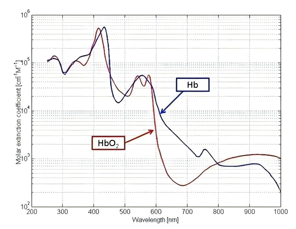
The molecular structure of hemoglobin will change depending on whether it carries oxygen, and the color will also change accordingly. We can use this property of hemoglobin molecules to distinguish the two. According to the colors of the two, we use near-infrared light as the light source to obtain the spectrum, build a transmission system, and use the SR50R17 spectrometer of JINSP to detect the absorbance of the hemoglobin sample, and we can get the following absorbance curve.

Biological tissue optics calls the wavelength range of 600–1000nm the biological spectral window. After using this wavelength band for light absorption experiments and drawing curves, it was found that HbO2 and Hb showed their own unique absorption spectra. At a wavelength of 660nm red light, Hb's absorption of light is significantly stronger than HbO2; while at a wavelength of 940nm infrared light, Hb's absorption of light is weaker than HbO2. Therefore, making good use of the different light absorption performance of hemoglobin under 940nm and 660nm light is the key to identifying the identity of HbO2 and Hb.
The following is a brief introduction to the experimental method of fine measurement:
We can mix PBS buffer, fat emulsion, melanin and hemoglobin in a certain proportion to make a human tissue phantom. Take fat emulsion and melanin of the same concentration and volume as when mixed and perform absorbance test to obtain the spectral data of the two groups of materials. Then change the concentration data of fat emulsion and melanin respectively to obtain multiple groups of data. According to the data, we analyze the influence of the two on the detection of hemoglobin. According to the concentration of the two in the human body, we process the spectral data obtained by measuring human tissue, and we can get a more accurate hemoglobin concentration, and then get a more accurate blood oxygen concentration.
In the above experimental process, we can use the SR50R17 spectrometer of JINSP to perform spectral analysis. If you are interested in Jinsp's products, please click the link below to contact us.
Post time: Aug-01-2024

